Obstetric ultrasound sound waves are employed to produce images of the foetus that lies inside a pregnant female's uterus. As ionising radiation is not involved in this procedure, it has no known harmful effects and is a preferred method used to monitor the growth and development of the unborn baby. Sometimes, a doppler ultrasound is done as a part of this examination, which involves an assessment of blood flow in the umbilical cord, placenta, or the foetus.
Before obstetric ultrasound, no special preparation is required. You should wear a two-piece outfit or a loose-fitting dress when going to the procedure room as only your lower abdominal area needs to be exposed. It is best to not wear jewellery to the procedure room.
Obstetrical ultrasound imaging is a noninvasive medical procedure used by physicians to diagnose and treat healthcare conditions that are related to the female reproductive organs. It is a painless, safe procedure during which images of the inside of the female reproductive organs are created with the help of sound waves.
Sometimes, a doppler ultrasound is done as a part of the obstetrical ultrasound. It is a special ultrasound technique which involves evaluation of the movement of materials inside the body. It allows the doctor to examine and evaluate the blood flow through the blood vessels in the body.
Having an accurate estimated due date is important for your baby so that the recommended tests can be performed at the same time. It is also important to know how far along you are if your baby is born prematurely or if you have failed to deliver the baby by your estimated due date and you are thinking about labor induction.
Most babies are delivered at around 38 weeks post-conception. Because many females ovulate and conceive around 2 weeks post their last periods, this is usually around 40 weeks as the beginning of their last menstruation. Therefore, people usually consider pregnancy to last for 40 weeks.
Females who have a regular 28-day cycle can calculate an estimated date of delivery for their baby by counting 40 weeks from the first day of last periods. This may not be too accurate and simple in other situations, suppose if you have irregular or long cycles, don’t remember your last date of period, or if you got pregnant while on contraceptives affecting your cycle.
If you think that you may require a dating scan to help estimate the due date of your baby, you must talk to your gynaecologist.
A dating scan is performed between 8-14 weeks of pregnancy when most babies of the same gestational age are almost of the same size. If your doctor thinks that you should have a dating scan and a nuchal translucency, they may recommend you to arrange it between 11-13 weeks, so that you can undergo both tests in a single ultrasound.
First-trimester screening is a combination of a special ultrasound and two blood tests that are employed to assess the risk of a pregnant woman carrying a baby with Edwards syndrome or Down syndrome. Performing and assessing them together and considering the age of the woman increases both the specificity and sensitivity of the screening results.
Pregnancy-associated plasma protein A (PAPP-A) is a protein that is produced by the growing placenta. The levels of this protein increases in the blood of a pregnant woman until delivery during a normal pregnancy. HCG is a hormone released in large quantities during pregnancy by the placenta. Either total hCG or free beta subunit can be used in first trimester screening. The levels of both generally rise fast in the blood of the pregnant woman for the first 8-10 weeks, then it decreases and stabilises at a lower level for the rest of the pregnancy.
Nuchal translucency is measured with the help of an ultrasound. The ultrasonographer quantifies the fluid collection between the spine and the skin at the nape of the neck of a foetus. It is a procedure requiring specially trained radiologists, proper alignment of the foetus, and careful measurement. It is not an ultrasound that is routinely performed and that is available at every health facility or hospital. If the results of first trimester screening are worrisome, diagnostic tests like chorionic villus sampling or amniocentesis may be recommended.
Blood is taken from a vein in the arm of the woman or collected from a finger stick. The nuchal translucency ultrasound is performed transabdominally (from outside the abdomen) or transvaginally (the probe may be introduced from the vagina).
An anatomy scan is also sometimes known as a 20-week ultrasound. It is a prenatal ultrasound done between 18-22 weeks of gestation. It is used to check on the physical development of the foetus and can help diagnose certain anatomical abnormalities and congenital disorders. Your doctor will use a 2D, 3D, or 4D ultrasound to take pictures of the foetus inside the uterus. The sonographer or ultrasound technician will take some measurements and ensure that the foetus is growing properly for its age.
The anatomy scan is used to take measurements of the foetal organs and body parts to ensure that the foetus is growing properly. The scan also looks for signs of structural issues with certain organs or congenital disabilities.
Some specific body parts of the foetus the doctor will examine include the brain, neck, spine, heart, kidneys and bladder, hands, fingers, feet and toes, arms and legs, chest and lungs, lips, chin, nose, eyes, and face, and stomach and intestines. The ultrasound technician will also check the umbilical cord to evaluate the blood flow, listen to the foetal heart rate for abnormal rhythms, check your ovaries, uterus, and cervix, and measure the amniotic fluid amount.
During this ultrasound, several images are taken and you will see that the ultrasonographer is drawing lines on the screen. This line serves as a ruler to document the sizes of the limbs and the organs. These measurements are compared against your due date. In some cases, you might hear that you are measuring behind or ahead of your due date. If the foetal measurements are within 10-14 days of the predicted due date, it is considered that the foetus is developing adequately.
Even though an anatomy scan doesn’t diagnose all the congenital conditions, it can help detect several serious conditions including down syndrome, trisomy 13, anencephaly, spina bifida, cleft lip, renal agenesis, congenital heart abnormalities, omphalocele, gastroschisis, diaphragmatic hernia, and skeletal dysplasia. It is important to note that the results of the scan are not a definitive diagnosis of any disorder. Instead, it is used as an indicator for determining whether or not further tests are needed. If your doctor is concerned with the scans, they will discuss the next steps with you.
A biophysical profile is an investigation that is usually performed after 32 weeks of pregnancy to evaluate the health of the foetus. It is often performed when the pregnancy is considered high risk due to certain health conditions or if you have aberrant test results. It helps answer questions about the well-being of your foetus. Following are the different areas the biophysical profile evaluate:
If your pregnancy goes beyond 40 weeks or if it is high risk, your pregnancy care provider may order a biophysical profile. They may also recommend a biophysical profile if you have any of the following conditions:
Biophysical profile has five parts - the first part of which is a non-stress test that evaluates the heart rate of the foetus in response to its contractions or movement. The other four parts are evaluated by an ultrasound. Those include looking at the foetal body movements, breathing movements, muscle tone such as flexing and extending the limbs, and measuring the amount of amniotic fluid.
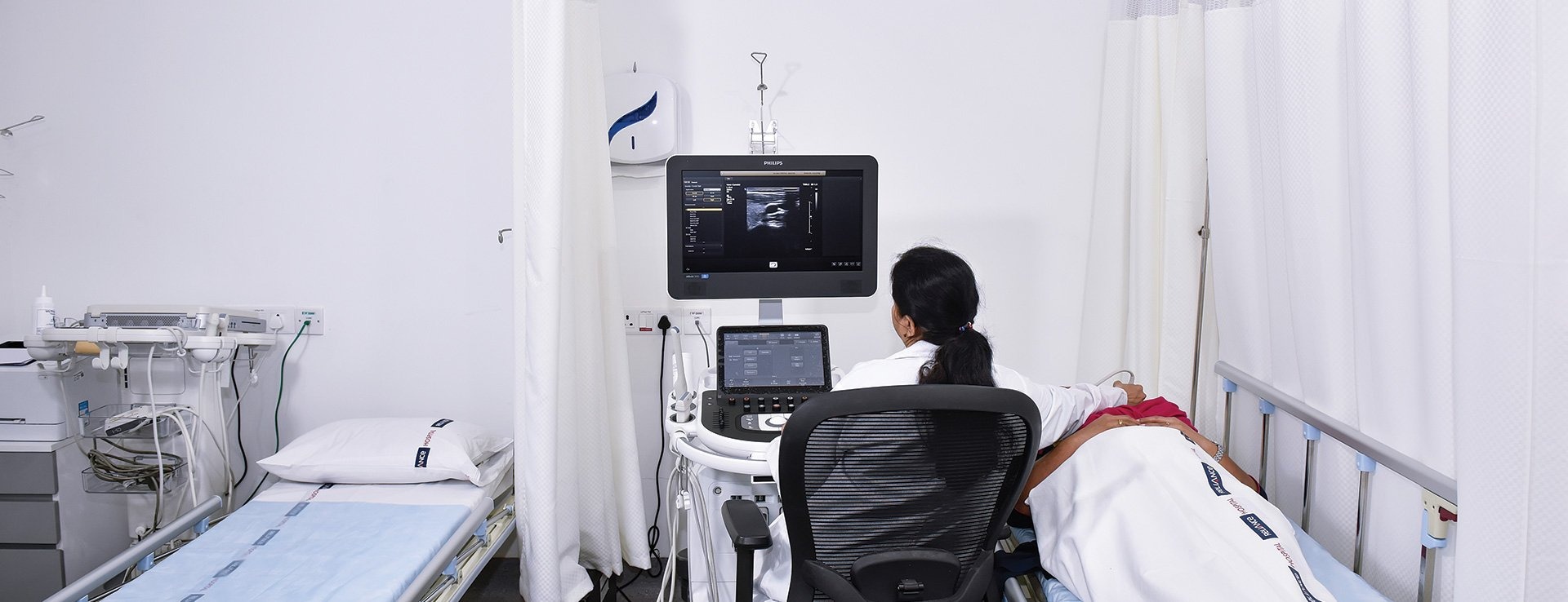
The sonography will first apply a hypoallergenic gel to the abdomen and move a transducer around the abdomen to collect images of the foetus. Then, he will move the transducer around the target area with moderate pressure to collect images. This pressure should not be painful. You must inform your doctor if you experience any discomfort. If you are facing problems holding your bladder or are in an uncomfortable position, you must talk to your doctor so that he makes some adjustments to make you more comfortable.
During the scan, the doctor will concentrate on completing your medical examination in a coherent and timely manner. So, you should try to save your questions for the end of the examination. Within some days, the radiologist will review the images and forward a detailed report to your doctor.
Some common uses of obstetrical ultrasound include establishing the existence of a living foetus, estimating the gestational age, diagnosis of congenital foetal anomalies, evaluating the placental and foetal position, determining the presence of multiple pregnancies, determining how much amniotic fluid is there around the baby, check for the cervical opening, and to evaluate the growth and well-being of the foetus. Some gynaecologists order a 3D ultrasound to get a detailed image of the foetus and determine if it is growing normally.
Following are some benefits of an obstetrical ultrasound:
Empathic and holistic maternity and newborn care is provided at the Department of Gynaecology and Obstetrics at Kokilaben Dhirubhai Ambani Hospital, Indore with an evidence-based approach toward normal pregnancy and delivery as well as their complications. Our team offers the most advanced obstetric ultrasound tests in Indore to help you achieve a normal, event-free pregnancy. Our maternity doctors work hard to render the process of childbirth as natural as possible while at the same time retaining the capacity and competency to manage all complications related to pregnancy.


