Diagnostic Radiology is the part of diagnostic medicine that is used by healthcare professionals to see the internal structures of the body. This helps them determine the cause of your symptoms, screen for diseases like cancer or heart disease, and monitor how well your body is responding to treatments.
Following are some of the most common diagnostic radiology tools used by healthcare professionals:
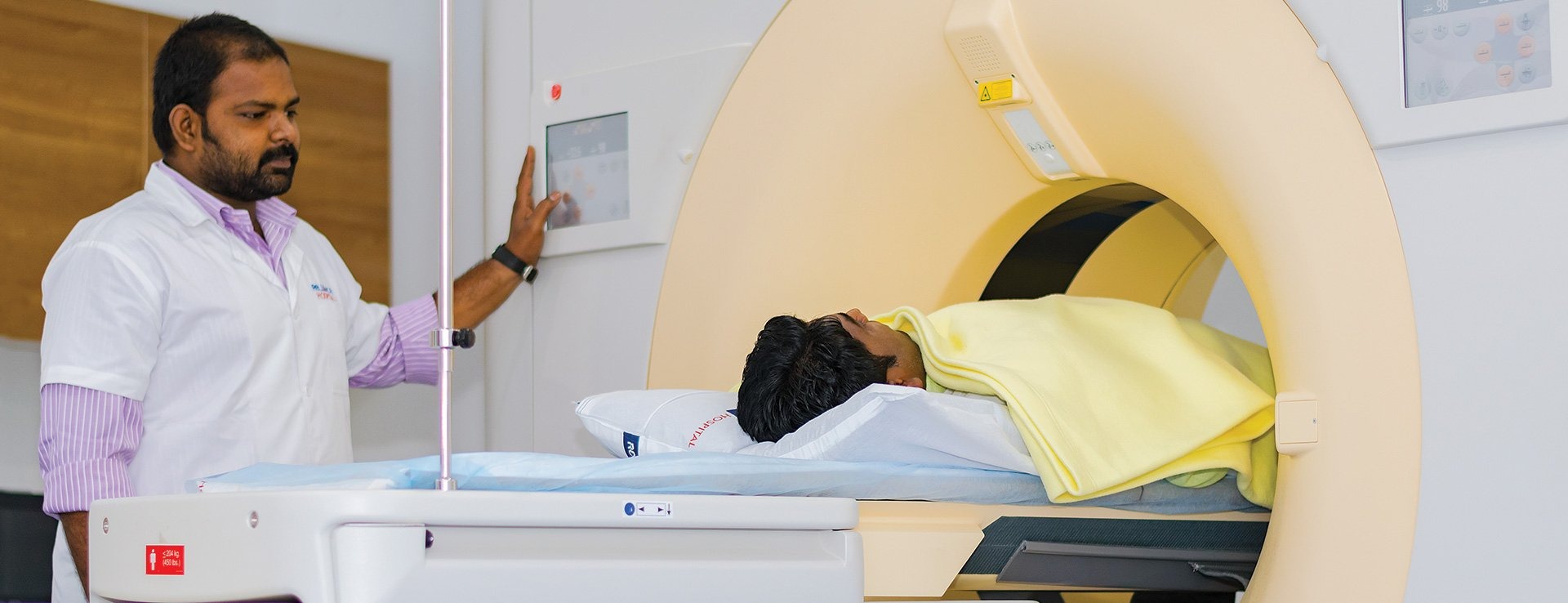
A CT scan combines a series of X-ray images taken from various angles around the body and employs a computer to produce cross-sectional images of the blood vessels, bones, and soft tissues inside the body. The images produced by CT scan provide more detailed information than those produced by plain X-rays.
A CT scan has many uses, however, it is most suitable for quick examination of patients with internal injuries from car accidents and other forms of trauma. Nearly all parts of the body can be visualised with the help of a CT scan to diagnose disorders or injuries.
Healthcare providers may recommend a CT scan to help:
Depending on the part of the body to be scanned, you may be asked to:
A special dye referred to as contrast material is required for certain CT scan procedures so that certain areas of the body are highlighted and being examined. The X-rays are blocked by contrast material and they appear white on images, which can help emphasise intestines, blood vessels, or other structures.
Contrast material might be given:
CT scans take only about thirty minutes and are generally painless.
During the procedure
CT scanners look like a huge doughnut standing on its side. You are directed to lie on a narrow, motorised table that slides into the tunnel through the opening. Pillows and straps may be used to help you stay in position. The table may be fitted with a special cradle holding your head still during a head scan.
While the table goes into the scanner, the X-ray tube and detectors rotate around you. Several images of thin slices of the body during each rotation. You may hear whirring and buzzing noises.
A technologist sitting in another room can hear and see you. You will be able to communicate with the technologist through intercom and he may ask you to hold your breath at intervals to prevent the blurring of the images.
After the procedure
You can return to your normal activities after the procedure. You may receive special instructions if contrast material has been injected. Some people may be asked to wait for a while before leaving to ensure that everything is fine. You will be asked to drink plenty of fluids after the exam to help your kidneys flush the contrast material from the body.
A magnetic resonance imaging (MRI) scan is a common diagnostic procedure that employs strong magnetic fields and radio waves to produce detailed pictures of the tissues and organs within the body. The scanner looks like a large tube with a table in the centre that lets the patient slide in. MRI is different from X-rays and CT scans as not harmful ionising radiation is used during the procedure.
MRI is used to examine the inside of the human body in a detailed way using non-invasive tools. Below are examples where an MRI scanner is used:
Before an MRI scan, very little preparation is required. On arrival at the hospital, the patient might be directed to wear a gown. As magnetic fields are used, it is important that no metallic objects are present in the scanner. So, the patient is asked to remove metallic accessories and jewellery that might interfere with the machine.
A person won’t be allowed to have an MRI scan if they have any metal inside the body including shrapnel, bullets, and other foreign bodies. This also includes medical devices like aneurysm clips, cochlear implants, and pacemakers. If you feel nervous or anxious in closed spaces, you should share it with your healthcare providers. They can help you with some medications prior to the procedure to make it more comfortable for you.
Sometimes, an intravenous contrast material is given to enhance the visibility of tissues relevant to the scan. A radiologist then takes the patient through the MRI scanning procedure and answers any potential questions that they may have. After the patient enters the scanning room, he is helped to lie down on the scanner table. The medical staff makes sure the patient is as comfortable as possible during the procedure. Headphones or earplugs may be provided to minimise the loud noises coming out of the scanner.
Once you are inside the scanner, a technician will communicate with you via the intercom and make you feel as comfortable as possible. The scan won’t be started until you are ready. It is important to stay absolutely still during the scan as any movement can disrupt the images. If you hear loud noises coming out of the scanner, know that it is perfectly normal. You might be asked to hold your breath at certain times depending on the images and the part of the body being scanned.
If you experience discomfort during the procedure, you can communicate it with the MRI technician via the intercom and request them to stop it or take necessary steps.
After the scan, the radiologist examines the pictures to check if any more are needed. If he is satisfied with the images, the procedure is stopped. After that, the radiologist prepares a report for the doctor who had ordered the scan.
Magnetic resonance angiography (MRA) refers to the MRI scan of the blood vessels. Unlike traditional angiography involving placement of a tube into the body, MRA is not an invasive procedure.
You may be directed to change into a hospital gown. You can also wear clothes like a t-shirt and a sweatpant that does not involve metal fasteners. Certain metals can cause blurry images, so it's best to avoid those.
You will be asked to lie at a narrow table, which slides into a large funnel-shaped scanner.
Some examinations involve a special contrast dye. The dye is usually given prior to the test via a vein (IV) in your forearm or arm. The dye allows the radiologist to visualise certain areas more clearly.
During the procedure, a healthcare professional will watch you from another room. It may take anywhere from one to two hours.
You may be directed not to drink or eat anything for upto 4-6 hours before the test. If you are afraid of confined spaces, you must share it with your healthcare provider. He may give you medicine that helps you feel more comfortable and less anxious during the procedure.
Before the test, tell your healthcare provider if you have artificial heart valve, brain aneurysm clips, inner ear implants, heart defibrillator or pacemaker, intrauterine device, insulin pump or chemotherapy port, neurostimulator, kidney disease or dialysis, vascular stent, recently placed artificial joints, or worked in sheet metal in the past. This is because the MRI machine works on the principle of strong magnetic fields and, therefore, metal objects are not allowed in the same room as the scanner when it’s working.
Avoid carrying items like watches, jewellery, credit cards, pens, hearing aids, pocket knives, hairpins, eyeglasses, pins, metal zippers, removable dental implants, and similar items.
MRA is used to visualise the blood vessels in all body parts. It may be performed on the heart, head, lungs, abdomen, legs, and kidneys. It may be used to detect or evaluate conditions like:
Mammography refers to the X-ray of the breast. Mammograms are used to look for signs of early breast cancer. These are of two types— diagnostic mammograms and screening mammograms.
A screening mammogram is one that is performed on females with no signs or symptoms of breast cancer. Regular screening helps reduce the number of deaths from breast cancer among women aged 40 and 74. This is because they can diagnose breast cancer early and start the treatment before it spreads and involves more tissues and nearby organs.
A diagnostic mammogram is performed on females who present with signs or symptoms of breast cancer, such as a lump or nipple discharge. Other signs include thickening of the skin of the breast or a change in breast shape or size. However, these signs can also occur due to a benign breast condition. Along with other tests, a mammogram allows the doctor to decide whether it is cancer or some other medical condition.
You will be directed to stand in front of an X-ray machine and the breast will be placed between two plastic plates that press and flatten it. This might be uncomfortable, however, it helps obtain a clear picture. Both the breasts are X-rayed from the front and the side. Then, a report is produced which the doctor evaluates for early signs of breast cancer or other problems.
If a mammogram comes out to be abnormal, it does not mean that you have breast cancer. You will require additional tests or exams before a doctor can tell for sure. You may also be referred to a surgeon or a breast specialist. However, it does not necessarily mean you have malignancy or require surgery.
X-rays are one of the commonest types of imaging tests that use a type of radiation called electromagnetic waves. Images of the internal structures of the body are created and the different parts of the body are shown in shades of black and white. This is because different amounts of radiation are absorbed by different tissues. Most X-rays are absorbed by calcium in bones, so they appear white on an X-ray. On the other hand, fat and other soft tissues absorb less radiation, so they appear grey. Lungs appear black as air inside them absorbs the least radiation.
X-ray techniques are used to examine different parts of the body.
Bones and teeth
Fractures and infections: Infections and fractures in the teeth and bones appear clearly on X-rays.
Chest
Abdomen
Abdomen
Different types of preparation are required for different types of X-rays. You should ask from your healthcare provider if you require any special preparation.
What to wear
Generally, the part that needs to be examined has to be exposed. You may be asked to change into a gown during the exam, depending on the area to be examined. You may also be asked to remove accessories like eyeglasses, jewellery, and any metal objects as they can show up on an X-ray and interfere with the results.
Contrast material
Sometimes, contrast agents like barium or iodine are given before an X-rat to help outline a specific area of the body on the X-ray image. You may be asked to swallow the contrast medium or receive it as an enema or an injection.
During the X-ray
You will be taken to the room where the X-ray machine is placed. The machine produces a safe level of radiation that travels through the body and collects images on a specialised plate. You won’t feel any pain during the procedure. A healthcare provider positions your body at certain angles to obtain the necessary views that may help in diagnosis. You are asked to stay still and sometimes hold your breath so that the image does not come out to be blurred. A simple X-ray procedure may take just a few minutes. More complex ones involving contrast media may take longer.
After the X-ray
After the procedure, you can go back to your daily activities. Routine X-rays generally don’t have any side effects. However, if contrast medium is given before the procedure, you must drink plenty of fluids to help the body eliminate it. If you experience pain, swelling, or redness after the procedure, you must talk to your healthcare provider.
Results
X-rays are digitally saved on a computer and the images can be visualised on the screen within minutes. Typically, a radiologist views and interprets the results and the report is then forwarded to the doctor who had ordered it.
Diagnostic ultrasound is a common imaging technique that employs sound waves to produce images of the internal structures of the body. The image provides vital information for diagnosing and directing treatment for a diverse range of diseases and conditions. Most ultrasounds are performed using an ultrasound machine outside the body. However, there are some techniques that involve placement of a small device inside the body.
Ultrasound is used for many reasons, including to:
Most ultrasound exams require almost no preparation at all. However, there are a few exceptions:
Clothing and personal items
Wear loose-fitted clothes to your ultrasound appointment. It’s a good idea to not wear any jewellery and leave valuables at home when coming to your appointment.
Before the procedure
Before your ultrasound starts, you may be directed to do the following:
The test reports are ready almost immediately and sent to the primary doctor who ordered the test.
The Department of Radiology at Kokilaben Dhirubhai Ambani Hospital, Indore, offers both diagnostic and interventional radiological services. Access the best diagnostic radiology services here. The department is equipped with state-of-the-art infrastructure and high-end diagnostic machines, and other medical instruments. We house a highly talented and dedicated team of medical professionals, including interventional radiologists, neuro radiologists, and allied staff. The team is highly experienced in dealing with such cases; therefore, they can decide the proper diagnostic and treatment plans for every patient in the best possible way.


