Interventional radiology involves image-guided procedures that radiologists perform using diagnostic imaging tools like MRI, ultrasound, and fluoroscopy to guide their procedures. Open and laparoscopic surgeries are avoided by most interventional procedures and minimally invasive options are favoured. Advantages of interventional radiology include greater comfort, lower costs, and faster recovery time. While it may not always prove to be the best procedure for your condition, it is generally more effective than conventional treatments.
Examples of interventional radiology procedures include:
The experts in the field of interventional radiology are known as interventional radiologists. They work closely with doctors of other departments and play a vital role on the treatment team.
Interventional radiologists perform a wide range of procedures, including:
An angiography is a diagnostic procedure that employs X-ray pictures to detect blockages in the blood vessels. An angiography test allows the doctor to see how the blood is flowing inside the blood vessels at particular locations in the body. An angiography is used to detect abnormalities in different organs of the body including heart, kidneys, neck, legs or other body parts to locate the origin of an arterial or venous issue.
Why is angiography performed?
An angiography is usually recommended by healthcare providers when they see signs of damaged, blocked, or abnormal blood vessels. An angiography helps them find out the source of the issue and extent of damage to the blood vessels. With the help of angiography, your healthcare provider can diagnose or treat conditions like peripheral artery disease, coronary artery disease, blood clot formation, atherosclerosis, and aneurysm.
How do I prepare for an angiography?
Before angiography, the healthcare provider may want to check your blood to find out how well it is able to form clots. They will also want to check if the kidneys are performing well. Following are some guidelines you can follow after midnight the night before the test:
Before deciding to discontinue any medication, especially anticoagulants and antiplatelet agents, always consult with your doctor.
If your healthcare provider approves:
Don’t take blood thinners like aspirin or any products containing aspirin. Take all your other medications as usual.
If you are diabetic, ask your doctor for instructions regarding when and if to take your insulin and/or oral medications.
Don’t eat anything post-midnight the night before the procedure. If general anaesthesia has to be administered during the procedure, don’t eat or drink anything post-midnight.
Consume only clear liquids for breakfast on the day of the angiogram. Such liquids include tea, clear broth, ginger ale, and black coffee.
What should I expect after an angiography?
Your doctor will remove the catheter and bandage the area of the skin which was punctured. The bandaged area is pressed for at least 15 minutes to stop or prevent bleeding. If the catheter is introduced into the leg, you will be directed to rest in bed for around 4-6 hours. This makes the incision less likely to bleed. Your doctor will assess and discuss at-home instructions with you before you are sent home.
Recovery after angiography
You will be able to go home the same day as the angiography procedure, even if you underwent angioplasty with stenting. Because anaesthesia had been given, someone needs to drive you home after the procedure. After you get home, you should not lift heavy stuff or bend or stoop for the next two days. This will prevent the incision from bleeding. Someone must stay with you overnight after the procedure. In some cases, you may need to spend the night at the hospital for recovery.
Also known as balloon angioplasty, this procedure involves opening of the arteries to let blood pass more easily through them. Doctors use this minimally invasive procedure in tight spots in arteries where plaque narrows the space inside an artery.
Who needs an angioplasty?
Those who have a history of heart attack or have coronary artery disease may need to undergo coronary angioplasty. The procedure is also used in other body parts that have blocked or narrowed arteries, such as the arms, neck, kidneys, legs, and pelvis. Angioplasty lets more blood flow through a narrowed or blocked artery, which means that the organ that the artery supplies receives a better blood supply after angioplasty.
What does angioplasty treat?
You will be directed to stop eating or drinking for a few hours prior to the angioplasty procedure. Depending on the situation, you can plan on your angioplasty procedure taking a half-hour to a few hours.
When you reach the hospital, you will be asked to change into a hospital gown. You must share your current and past medication history with your healthcare provider including any history of allergies you might have. The doctor will introduce an intravenous line into your arm so that medicine can be given to relax you. You will still be able to respond to the question your healthcare provider will ask you. You will also receive medication in your IV line to prevent your body from producing blood clots.
Advantages of angioplasty include:
You will be required to stay at the hospital for several hours or even overnight to recover from the procedure. Your doctor will let you know what medicines you need to take at what time and how active you can be after your angioplasty. You will require someone to drive you home from the hospital as you have been given anaesthesia.
Get rest at home and maintain adequate fluid intake: Don’t exert yourself too much for the next twenty-four hours. You may be required to take medicines such as blood-thinners after the procedure. Make sure that you take these medications as prescribed by your healthcare provider. Don’t miss any doses. If you feel like you need to stop taking these medications, talk with your healthcare provider before you actually decide to stop taking them.
When can I go back to work or drive?
You will be able to go back to work and drive usually a week after the coronary angioplasty. For other types of angioplasty, the time may be shorter as the recovery is quicker. Your doctor will let you know the specific instructions that you need to follow according to your procedure and overall condition.
It is a minimally invasive technique used to block or close a specific blood vessel. Embolisation is usually performed as an elective procedure. Some of the cases are emergencies and need to be performed right away.
How is embolization helpful?
Embolization provides temporary or permanent relief for a range of conditions by:
What conditions does embolization treat?
Embolization can be used for the treatment of conditions affecting nearly every body part. The procedure can be helpful for patients having brain aneurysms, arteriovenous malformations, frequent nosebleeds, cancers and tumours causing bleeding, long menstrual periods or ones with excessive bleeding, gastrointestinal bleeding from conditions like diverticulosis and stomach ulcers, overactive spleen, traumatic injuries affecting certain organs like liver, spleen, and lungs, uterine fibroids, retroperitoneal hematoma (bleeding in the space behind the lining of the abdominal wall, vascular malformations including abnormal connections between the veins and arteries, and varicocele (swollen veins in the scrotum).
How does an embolization procedure work?
During embolisation, tiny objects or particles known as embolic agents to halt the flow of blood. The doctor delivers the agents using thin, long tubes. These tubes known as catheters are inserted through a puncture in the skin and then followed through the path of the blood vessel to reach the treatment area. Instruments attached to the tip of the catheter make it possible to carry out the procedure.
What types of embolic agents are used?
The type of embolic agent used depends on your medical needs and the blood vessel being treated.
The types of embolic agents include:
What are the different techniques used in embolization?
Embolization is of many types, including:
What are the advantages of embolization?
Embolization offers many advantages, including:
Gastrostomy tubes: A feeding tube is introduced into the stomach if you can’t take food by oral route.
Intravascular ultrasound (also known as coronary intravascular ultrasound, intravascular echocardiography, endovascular ultrasound) is a technique that employs sound waves to assess soft tissue. Small instruments are used to collect real-time image from inside the blood vessels. The arteries and veins near the heart are evaluated by an intravascular ultrasound. Other blood vessels can also be tested.
How does intravascular ultrasound work?
Intravascular ultrasound uses a small tube called catheter to access and evaluate tissues of a blood vessel. A doctor introduces the catheter through an incision, typically in the groin to reach the assessment area.
The catheter is attached to an ultrasound probe at the tip that emits high-frequency sound waves. These sound waves create echoes by bouncing off the walls of the blood vessels and then these are converted into real-time images.
How can an intravascular ultrasound procedure help me?
You may benefit from an intravascular ultrasound procedure if healthcare providers suspect narrowing or blockages in blood vessels. The procedure is used in the management of life-threatening conditions including pulmonary embolism, heart attack, and stroke.
IVUS helps healthcare providers:
How is an intravascular ultrasound performed?
If your doctor is assessing your coronary arteries, intravascular ultrasound is typically performed as a part of cardiac catheterization. This procedure employs a catheter to check the cardiac function. Combining the two tests makes it possible to check additional aspects of cardiac health in a single procedure.
If the area of assessment is not near the heart, intravascular ultrasound is performed as a standalone procedure. It is often employed to check for blood clots in the veins and peripheral artery disease in the lower leg arteries.
Who performs intravascular ultrasound?
Intravascular ultrasound is performed by an interventional cardiologist.
What happens during an intravascular ultrasound procedure?
You will first be asked to lie down on a table and receive medication to help you relax. You may be conscious and awake but will not feel uncomfortable during the procedure.
A healthcare professional will clean the skin near the site of incision. Then, they inject a numbing medication to desensitise the area. The vascular surgeon or interventional cardiologist slides a plastic sheath into the incision, making it easier to introduce the catheter and advance it. After the catheter reaches the right spot, they use the probe to capture pictures. Once the procedure is over, the healthcare professional removes the sheath and the catheter. Usually, stitches are not necessary to close the incision and infection is prevented by a surgical dressing.
What are the benefits of IVUS?
Intravascular ultrasound procedure is used by healthcare providers to detect any narrowing or blockage in the blood vessels. Additional benefits include:
Foreign body removal refers to removal of foreign objects that have entered accidentally into the body. Foreign agents can be introduced into different body parts including eye, ear, finger, nose, foot, leg, skin, stomach, breathing tract and more. The technique employed to remove the substance depends on the substance and its location in the body.
Foreign bodies may occur if something has been inserted or ingested by the patient or someone else into the body. They can occur because of an accident. In some cases, ingested agents can pass via the digestive tract naturally and without complications, however, some patients may require assistance to remove the foreign body. Foreign bodies may also be required to be removed by an emergency room doctor if they cause pain, or are in the skin or eye.
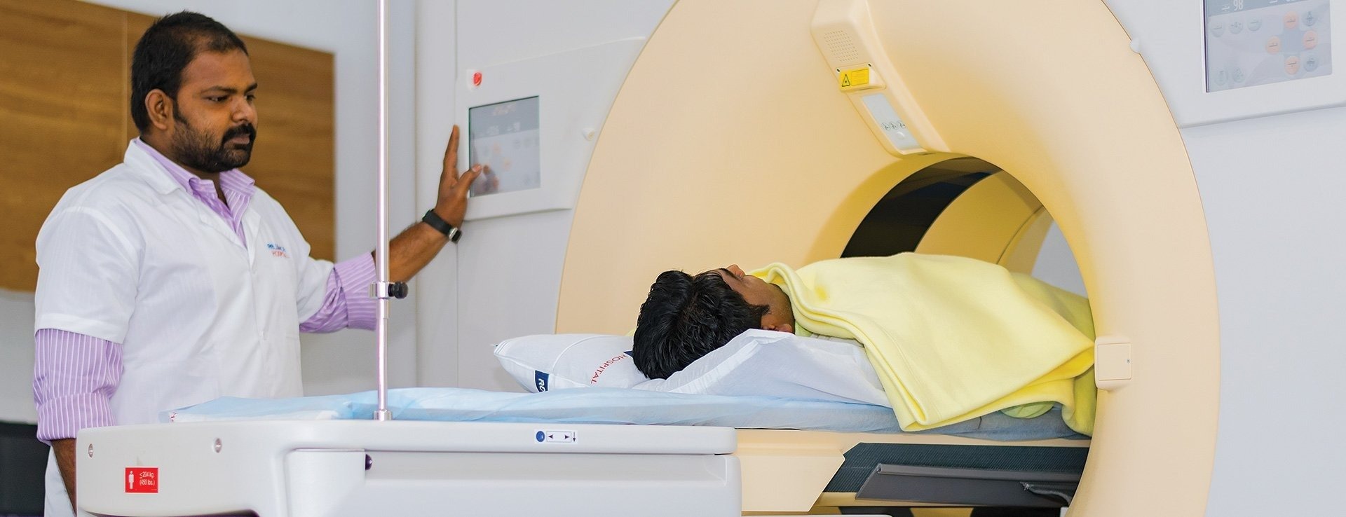
The Department of Radiology at Kokilaben Dhirubhai Ambani Hospital, Indore, offers both diagnostic and interventional radiological services. Consult the best interventional radiologists in Indore here.The department is equipped with state-of-the-art infrastructure and high-end diagnostic machines, and other medical instruments. We house a highly talented and dedicated team of medical professionals, including interventional radiologists and neuroradiologists in Indore. All of them have years of experience in dealing with such cases, therefore, they can decide the proper diagnostic and treatment plans for every patient in the best possible way.


